
Causes of pain following ankle sprain
Ankle sprains vary in severity. While the majority of ‘simple’ ankle sprains go onto a good recovery, there are a substantial subset of patients with more ‘complex’ injuries that have ongoing pain following ankle sprain.
At the time of the injury, it is possible for damage to occur to the ankle joint itself, as well as the surrounding soft tissues and tendons.
Pain following ankle sprain is often associated with ongoing swelling and a loss of confidence in the stability of the ankle. Depending on the location of the pain, various pathologies need to be considered.

Pain at the front of the ankle (Anterior ankle pain)
This is a common area to experience ongoing pain following ankle sprain. Pain in this region is likely due to either joint surface (chondral/articular surface) damage or associated ‘impingement’ from soft tissue scarring.
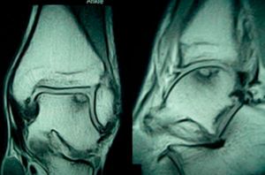
Talus OCD (cartilage damage)
Articular cartilage damage may include displacement of a section of the joint surface of the ankle. The ‘Talus’ is a large bone of the ankle that articulates with the lower end of the tibia (shin bone) and fibula.
Cartilage damage can occur in the anterolateral (front/outside) part of the talus, following a twisting injury. Impaction occurs between the talus and the lower end of the fibula, resulting in loss of cartilage in this area.
Patients will present with ongoing pain at the front and outer aspects of the ankle joint, with associated swelling. ‘Catching and clicking’ of the joint is often experienced. Patients experience difficulty walking on uneven ground.
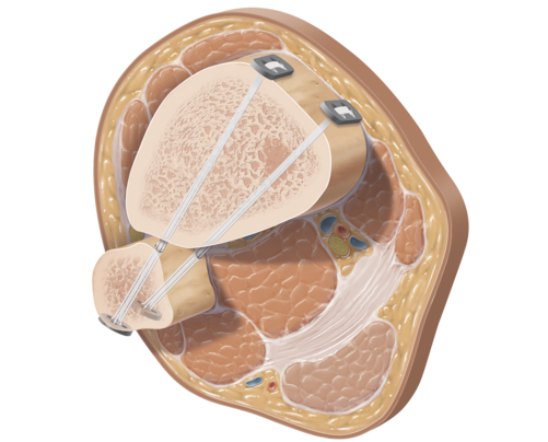
X-rays and advanced imaging (MRI scan) will confirm the severity of the cartilage damage, and a tailored management plan is discussed. A keyhole ankle arthroscopy is frequently required, and then the cartilage damage needs to be addressed.

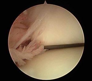
Loose bodies inside the ankle joint
During a high energy ankle sprain, cartilage impaction can result in the formation of loose bodies that become entrapped within the ankle joint. These ‘bodies’ can increase in size once detached. Patient will typically have ongoing pain and the sensation of something ‘catching’ inside the ankle joint. The loose bodies may move around inside the joint causing intermittent ‘locking’ of ankle movement.
Following evaluation with an x-ray and MRI scan, keyhole ankle arthroscopy is performed to remove these ‘loose bodies’ and evaluate the cartilage surface of the ankle joint.
Soft tissue or Bony impingement
Anterior ankle impingement is characterised by pain at the front of the ankle joint during activities that result in ‘dorsiflexion’ of the ankle. Patients typically have difficulty with walking upstairs, landing from a jump, and pushing the brake in the car. Impingement occurs due to compression of bony or soft tissue structures during these activities.
Soft tissue scarring and impingement can cause ongoing pain following ankle sprain.
The majority of these conditions respond well to a keyhole arthroscopic ‘decompressions’.
Syndesmosis Injury (‘High’ ankle sprain)
The ankle syndesmosis is a ‘fibrous joint’ between the lower end of the tibia (shin bone) and fibula. Syndesmotic injuries are often referred to as a ‘High ankle sprain’ as the torn ligaments of the syndesmosis, lie above the more commonly injured lateral ankle ligaments.
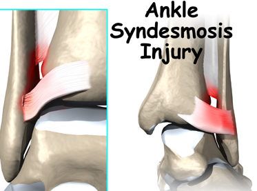
This injury often occurs when the foot is planted on the ground, and an excessive outward twisting of the foot occurs.
The pain is often located just above the level of the ankle joint, and is made worse with external rotation of the foot. Syndesmotic injuries can also occur in the setting of an ankle fracture, which require an operation.
An early diagnosis of a syndesmosis injury is important, as the management differs from a ‘simple’ ankle sprain. Depending on the severity of the injury, surgery to stabilise this ‘joint’ may be required.
Pain at the back of the ankle (Posterior ankle pain)
This is a less common region to experience ongoing pain. This typically represents ‘posterior ankle impingement’ that can occur due to the abutment of soft tissue or excessive bone (Os Trigonum).
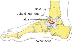
Pain on the inner side of the ankle (Medial ankle pain)
Depending on the mechanism of injury, ongoing pain on the inside of the ankle joint may represent a missed bony injury (sustentaculum tali fracture), deltoid ligament rupture, or an injury to the talus articular cartilage on the inner (medial) side.
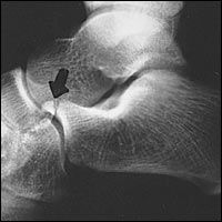
Sustentaculum Tali fracture
The ‘Sustentaculum Tali’ is a prominence of bone that is part of the calcaneus (Heel bone). It is positioned on the inside of the foot, and tendons to the toes run above and below this section of bone.
This diagnosis is easily missed and requires a high index of suspicion. A CT scan helps confirm the diagnosis and management may require screw fixation for optimal outcome.
Deltoid ligament Tear
The deltoid ligament is a broad fan shaped ligament that lies on the medial (inner) side of the ankle. Injury to this ligament occurs with excessive ‘external rotation’ of the foot. It can be seen in isolation, but often is in combination with a lateral sided injury – Ligamentous of fracture.
Patients experience ongoing inner side of foot pain with associated swelling and weakness.
The diagnosis is confirmed with an x-ray and MRI scan. Depending on the severity of the injury, surgery may be required.
Talus OCD (cartilage damage)
While cartilage damage more commonly affects the lateral (outer) side of the talus, following an injury, it is possible for the medial (inner) talus cartilage to be damaged also.
Treatment of these ‘lesions’ can be challenging and requires a detailed assessment first.
Pain on the outer side of the ankle (Lateral ankle pain)
This is the most common location for pain following ankle sprain. There are many causes that require a thorough assessment and management plan.
Ankle synovitis
Ankle synovitis is inflammation of the soft tissue lining of the ankle joint. This can occur following an ankle sprain, with pain and swelling felt just in front of the lateral malleolus (end of the fibula). If this pain fails to settle, an ankle arthroscopy may be performed to remove the inflamed tissue.
Soft Tissue scarring within the ankle joint
The typical ankle sprain results in tearing to the lateral ankle ligaments. Scarring of these ligaments can result in a condition termed ‘fibrosis’. This scarring can cause pain on the outer side of the ankle joint, and responds well to an arthroscopic ‘debridement‘ if it fails to settle.
Missed bony fracture
Ongoing lateral (outer) sided ankle pain, may be the result of a missed fracture. The most common locations for this include:
- Anterior process of Calcaneus
This is a part of the calcaneus (heel bone) that articulates with a small bone in the foot called the cuboid. Fracture to this areas is typically due to an ‘avulsion’ of the ‘bifurcate ligament’.
- Lateral process of talus
This is a part of the talus that lies just below the tip of the fibula, on the outside of the foot. X-rays commonly ‘miss’ the fracture, and a high index of suspicion is required to make the diagnosis.
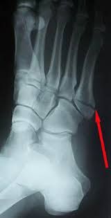
- Base of 5th metatarsal fracture
The 5th metatarsal is a long bone on the outside of the foot. The peroneus brevis tendon attaches to the base of the metatarsal, as well as part of the plantar fascia. An ‘inversion’ injury may result in an ‘avulsion’ fracture to this region, and the diagnosis is confirmed with plain x-rays. Depending on the specific location of the fracture, screw fixation may be required.
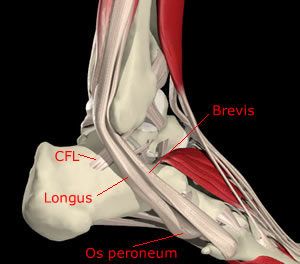
Peroneal tendon tear/inflammation
The peroneal tendons run down the outside of the leg and behind the lower end of the fibula. They are vital in providing stability to the ankle joint. The peroneal tendons (especially the peroneus brevis) may be torn as a result of the ankle injury. Ongoing pain on the outside of the ankle (behind the lower fibula) and swelling will occur. An MRI scan confirms the diagnosis and surgical repair is often required.
Do you experience any of the following?
Frequent Ankle Sprains
Pain in your ankle
Swelling around your ankle